Time:2013-01-17
On Jan 9th, 2013, a recent study from Dr. Mingsha Zhang’s lab at the Institute of Neuroscience, Chinese Academy of Sciences, was published online in the Journal of Neuroscience. In this study, Dr. Zhang and his colleagues investigated the neuronal control of saccadic eye movements through recording single neuron’s activity from the posterior parietal cortex of non-human primates. Based on both behavioral and neuronal data, they show that the activity of many neurons in the monkey’s posterior parietal cortex is highly correlated with the generation of express and regular saccades. Their findings challenge the classical view of express saccades being purely controlled subcortically, sheding new insight in understanding the neuronal control of saccade generation.
Saccadic eye movements are of great importance for primates to direct their eyes to objects of interests. On average, primates may make 3 saccades per second. When saccades are directed to a predicted location, some of the saccades occur with very short saccade reaction time (SRT) and these saccades are named express saccades. To study the neuronal mechanism of express saccades, Dr. Zhang’s group recorded single neuron’s activity in the lateral intraparietal cortex (LIP), a subregion of the posterior parietal cortex, of macaque monkeys during a gap saccade task. In gap tasks, rather than target onset occurring simultaneously with fixation spot offset, a delay (gap) precedes target onset. They found that, as compared to the no-gap task, 50% of LIP neurons increased their activity during the gap interval in the gap task. This pretarget preparatory activity might play a role in the occurrence of short latency express saccades. Furthermore, the persistent neurons, a subset of LIP neurons, showed enhanced activity during express saccades but not during regular saccades, from the beginning of initial fixation until target onset. Such increase is spatially selective and is limited to the neuron’s response field. However, the visual neurons did not show any significant differences in response between express saccade and regular saccade trials. These results indicate that the enhanced preparatory activity plays a crucial role in determination of the express saccade generation. This study provide the first electrophysiological evidence that parietal cortical neuronal activity is selective for express saccades and support the posterior pathway hypothesis which implicates the posterior parietal cortex (PPC) in express saccade generation.
The experiments were carried out by two graduate students Mo Chen and Yu Liu, and visiting scientist Linyu Wei. This study was supported by 973 program and Pujiang Program.
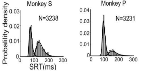
During gap task, the SRT distributions usually show bimodal profiles. Some saccades generate with very short latency forming the first mode, named as express saccades.
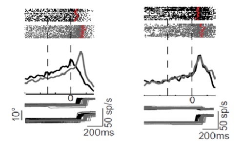
A group of LIP neurons show significant greater activity during express saccades than that during regular saccades as shown in the left panel whereas others do not show significant difference between two types of trials as shown in the right panel.
 附件下载:
附件下载: 
