Time:2014-01-27
On Jan. 26th, Dr. Xiang Yu’s research group at the Institute of Neuroscience, Chinese Academy of Sciences, published a research article entitled "Oxytocin mediates early experience-dependent crossmodal plasticity in sensory cortices" in the journal Nature Neuroscience as Advance Online publication.
During the early postnatal period, natural sensory experience is important for the development of the nervous system. Deprivation in one sense not only reduces responses in the correspondent cortical region, but also crossmodally affects other brain regions. Although the crossmodal effects of sensory experience have been investigated in adult individuals at both electrophysiological and behavioral levels, little is known about the mechanism of crossmodal plasticity during early development. Recently, a research team led by Dr. Xiang Yu at the Institute of Neuroscience, Chinese Academy of Sciences, identified a novel form of plasticity in neonatal mice, where early sensory experience crossmodally regulates development of all sensory cortices via oxytocin signaling.
In this study, Dr. Yu's group reported that whisker deprivation or dark rearing from birth effectively reduced the frequency of miniature excitatory postsynaptic currents (mEPSCs) and spontaneous firing rates in layer II/III pyramidal neurons, both in the correspondent sensory cortex, and crossmodally in other sensory cortices. Sensory experience regulated the synthesis and secretion of the neuropeptide oxytocin from the hypothalamus, as well as its cortical level. Importantly, in vivo oxytocin application significantly increased excitatory synaptic transmission and rescued the effect of sensory deprivation in multiple sensory cortices. Conversely, sequestering endogenous oxytocin or interfering with its signaling reduced excitatory synaptic transmission. Finally, increasing natural sensory stimulation through environmental enrichment increased oxytocin level and attenuated the effect of sensory deprivation.
This work identified a novel form of experience-dependent crossmodal plasticity in sensory cortices during early development, in which oxytocin functions as a key mediator, at a much earlier developmental time point than its previously described roles in mediating social emotional behaviors. The link between sensory experience and oxytocin signaling may be particularly relevant to autism research, where part of the hotly debated therapeutic role of oxytocin may be mediated through its effect on sensory cortices, as hypersensitivity or hyposensitivity to sensory inputs is reported to be prevalent in children with autism spectrum disorders.
This work was carried out by graduate student Jingjing Zheng, postdoctal fellow Shujing Li and colleagues under the supervision of Dr. Xiang Yu, in collaboration with Dr. Haishan Yao. The study was completed at the Institute of Neuroscience and State Key Laboratory of Neuroscience, Shanghai Institutes for Biological Sciences, Chinese Academy of Neurosciences, and supported by grants from the Ministry of Science and Technology and the National Natural Science Foundation of China.
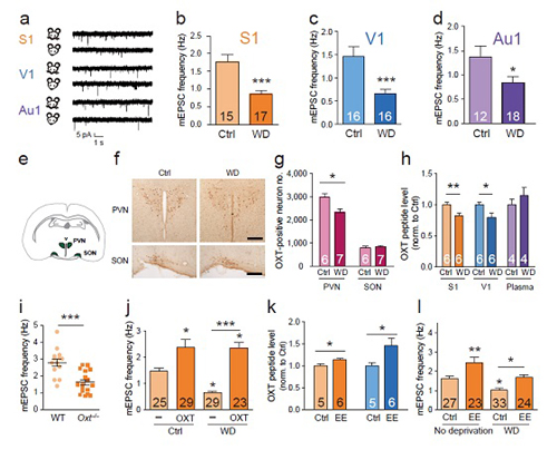
Figure legend: the effects of sensory experience and oxytocin in regulating cortical development.
(a) Representative miniature post-synaptic current (mEPSC) recordings for conditions as indicated from the primary somatosensory cortex (S1), primary visual cortex (V1) and primary auditory cortex (Au1).
(b) Whisker-deprivation (WD) significantly reduced mEPSC frequencies in S1.
(c) WD significantly reduced mEPSC frequencies in V1.
(d) WD significantly reduced mEPSC frequencies in Au1.
(e) Illustration of the location of the paraventricular (PVN) and supraoptic (SON) nuclei, both in the mouse hypothalamus, in a coronal brain slice.
(f) Representative images of oxytocin-positive neurons in the PVN and SON of P14 mice, conditions as indicated; scale bar = 200 μm.
(g) WD reduced the number of oxytocin-positive neurons in the PVN, but not SON.
(h) WD significantly reduced oxytocin peptide level in S1 and V1, but not blood plasma.
(i) Oxytocin knockout mice showed reduced mEPSC frequencies in S1.
(j) Oxytocin injection increased mEPSC frequencies and rescued the effects of whisker-deprivation in S1.
(k) Environmental enrichment (EE) significantly increased oxytocin peptide level in S1 and V1.
(l) EE increased mEPSC frequencies and rescued the effects of whisker–deprivation in S1.
 附件下载:
附件下载: 
