Time:2014-09-22
Emotions color our lives and drive behaviors. Most emotions can be categorized along two dimensions: valence (value), ranging from negative to positive, and salience (arousal, intensity), ranging from weak to strong. Although great advances have been made toward understanding the sensory representations of the external world, how different emotional valences are represented in the brain has remained largely elusive.
To extract the valence representation of an emotion in the whole brain, Dr. HU Hailan’s lab devised a strategy to compare the activity patterns evoked by two emotional stimuli with contrasting valence (morphine and foot shock) in the same mouse brain, by modifying a double-labeling technique based on the distinct induction time course of the mRNA and protein signals of the IEG gene c-fos. c-fos expression is generally low throughout the limbic nervous system in resting animals; however, following neuronal activation, c-fos mRNA accumulates in minutes and decays in a couple hours, whereas its protein product appears slower and lasts much longer. Thus, when two stimuli are sequentially applied at an appropriate interval, neural circuits activated by the two stimuli can be represented by the c-fos protein and mRNA signals respectively, which in turn can be revealed by dual labeling using fluorescence immunohistochemistry (FIHC) and fluorescence in situ hybridization (FISH).
Using the TAI-FISH dual labeling technique, they examined the c-fos activation patterns elicited by morphine and foot shock in the same mouse brain. They found that morphine and foot shock evoked largely distinct neural ensembles throughout the limbic forebrain, despite activating many of the same regions. As morphine and foot shock have different sensory properties, they also introduced additional pairs of emotional stimuli, including chocolate and restraint stress. In the nucleus accumbens (NAc), rewarding and aversive stimulus pairs (morphine/foot shock, morphine/restraint) recruited spatially intermingled neurons, whereas stimulus pairs of similar valence (morphine/chocolate, foot shock/restraint) activated largely overlapping neural groups, suggesting the existence of a functional valence map. These results provide insights into the internal representation of emotional valences and the functional organization of NAc. In addition, with the new amplification step allowing better separation of IEG mRNA and protein signals, TAI-FISH provides a much-needed molecular imaging technique for mapping the neural representations of two distinct behaviors across the whole brain at single-cell resolution.
This research entitled “Visualizing an emotional valence map in the limbic forebrain by TAI-FISH” was published online in Nature Neuroscience on September 21, 2014. This work was mainly carried out by graduate students XIU Jianbo and ZHANG Qi under the supervision of Dr. HU Hailan, in collaboration with graduate students ZHOU Tao, ZHOU Tingting, and research assistant CHEN Yang. The work was supported by the 973 Project of the Ministry of Science and Technology of China, the Outstanding Youth Program, the Strategic Priority Research Program of the Chinese Academy of Sciences.
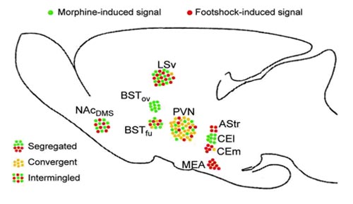
Figure 1 Latest study from Dr. HU Hailan’s lab unraveled an emotional valence map in the forebrain. Summary of patterns of interaction between neural representations of morphine and foot shock in different regions of the limbic forebrain, as revealed by TAI-FISH.
Figure 2 The intermingled pattern of neurons encoding opposite valence in nucleus accumbens.
 附件下载:
附件下载: 
