Time:2015-06-09
A recent study published on the Journal of Cell Biology demonstrated that the neuron migration is controlled by three highly dynamic contraction centers (CCs) which are regulated by guidance cues. This work was performed mainly by graduate student JIANG Jian in the laboratory of POO Mu-ming, at the Institute of Neuroscience, Shanghai Institutes for Biological Sciences, Chinese Academy of Sciences.
Migration of neurons from their birthplace to the designated areas is a critical step in the development of brain architecture. Although much progress has been made in our understanding of the cellular and molecular basis of neuronal migration, the biomechanical aspects of neuronal migration remains largely unclear. In this study, researchers directly measured the traction force of cerebellar granule cells (GCs) using traction force microscopy and made three major findings.
First, three distinct and independently fluctuating CCs exist in each GC, and the net action of CC activity drives soma translocation. Second, both F-actin and MTs contribute to the traction force generation, through myosin-IIa€“dependent processes. Third, the attractive and repulsive guidance cues, BDNF and Slit2, respectively, exert their influence on GC migration by altering spatiotemporal dynamics of CC activities at LPs and TPs. These results resolved the previous controversy over the location of force generation that drives neuronal migration, and showed that CC-generated pulling rather than pushing is responsible for soma translocation.
This research entitled "Spatiotemporal dynamics of traction forces showthree contraction centers in migratory neurons " was published in the Journal of Cell Biolog on June 8th, 2015. This work was supported by grants from Ministry of Science and Technology and the Chinese Academyof Sciences.
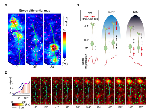
Figure legend: (a) Stress differential maps showed three distinct contraction centers (dashed circles) in cerebellar granule neurons (green lines). (b)The imbalance of traction force drove soma translocation. Left, Soma translocation with time. Two asterisks indicate the time interval shown on theright. Right, Stress maps at a continuous series of time points corresponding to the time interval marked by the arrow on the left. The cyan linesrepresent the cell outlines. (c) A model for traction force regulation during neuron migration.
 附件下载:
附件下载: 
