Time:2015-11-20
Dopamine release can be evoked by a hedonic olfactory stimulus, monitored in real time, and traced to its circuit origin in the transparent brain of zebrafish larvae, as reported on Nov. 18th in the Journal of Neuroscience. Using carbon fiber microelectrodes, this work demonstrated for the first time that sensory-evoked dopamine release can be successfully monitored in the brain of an awake small organism. By elucidating the circuit origin of the dopamine release, the potential application of this method in the dissection of the circuit mechanisms underlying dopaminergic neuromodulation was demonstrated.
Dopamine plays crucial roles in a broad spectrum of brain functions, and neural circuit mechanisms underlying dopaminergic regulation have been intensively studied in the past decade. As larval zebrafish has relative simple and highly conserved dopaminergic circuitry, it can serve as an ideal vertebrate animal model to tackle this issue at a whole-brain scale. For this purpose, it is important to develop methods for monitoring endogenous dopamine release in intact larval zebrafish. Being a gold standard in monitoring dopamine release in mammals, electrochemical recording has provided primary understanding on how phasic dopaminergic signaling participates in various neural functions. However, since larval zebrafish have much fewer dopaminergic neurons, several orders lower than mammals previously targeted with C-fiber microelectrodes, it is challenging to monitor dopamine release in such a small organism. With refined C-fiber microelectrodes, the authors succeeded in monitoring food-evoked dopamine release in the optic tectum of zebrafish larvae, with an amperometric current as small as several to dozens of picoamperes. Furthermore, with its tiny and transparent brain, zebrafish larva provides easy access to dopaminergic nuclei and their interconnected brain areas. By combining dopamine release monitoring, calcium imaging and laser ablation, the detected dopamine release was determined to be from pretectal dopaminergic neurons, demonstrating the potential of zebrafish larvae as a working model for thoroughly dissecting the functions and underlying mechanisms of dopaminergic systems.
This work entitled "Amperometric Monitoring of Sensory-Evoked Dopamine Release in Awake Larval Zebrafish" was carried out by Dr. SHANG Chunfeng and collaborators under the supervision of Dr. DU Jiulin at the Institute of Neuroscience, Chinese Academy of Sciences, in collaboration with Dr. ZHOU Zhuan at the Institute of Molecular Medicine, Peking University. It was supported by grants from the Chinese Academy of Sciences, the Ministry of Science and Technology of China, the Natural Science Foundation of China, and the Science and Technology Commission of Shanghai.
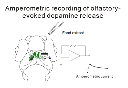
A carbon fiber (C-fiber) microelectrode was placed in the neuropil of the optic tectum (OT) and food extract was applied pneumatically at the ipsilateral nostril through a glass micropipette. Dopaminergic neurons (in green) at the pretectum were activated by the olfactory stimuli and released dopamine to the OT, resulting in an amperometric current response recorded with the C-fiber microelectrode.
 附件下载:
附件下载: 
