Time:2023-05-22
A new study published in Nature Neuroscience provides a comprehensive survey of the diversity and morphological types of dendrites of projection neurons in prefrontal cortex, by reconstructing and analyzing the complete morphology of dendrites and axons of nearly 2,000 single neurons in prefrontal cortex. This work reveals the relationship between dendritic morphology and axonal projections, and reconstructs the connection network between projection neuron types in prefrontal cortex. This work provides important clues for the in-depth study of the function of prefrontal cortex, basis for neuron simulation, and inspiration for artificial intelligence.
This study was performed by Dr. YAN Jun’s research group at the Institute of Neuroscience, Center for Excellence in Brain Science and Intelligence Technology of the Chinese Academy of Sciences.
As the basic structural compartments of neurons, dendrites and axons are responsible for integrating and transmitting neural information respectively. Studying the diversity of dendritic morphology and its relationship with axonal projections is important for understanding the rules of information processing in the neural network. The dendrites of cortical projection neurons display elaborate morphology and has been used as one of the important features for neuronal classification.
Previous studies on dendrites mainly reconstruct the dendritic morphology of cortical neurons on brain slices. However, this approach has missed the three-dimensional spatial locations of neurons, and the reconstructed dendritic morphology was often incomplete, making it difficult to uncover the full diversity of dendritic morphology.
In addition, to investigate the relationship between dendritic morphology and axonal projections, previous studies mainly injected retrograde tracers in downstream target areas. However, cortical projection neurons often have numerous projection target areas, and the use of retrograde labeling requires a priori knowledge of target area information, as a result the complete axonal projection information often cannot be obtained.
Therefore, a systematic study of the diversity of dendritic morphology and its relationship with axonal projections requires the mapping and analysis of the complete dendritic and axonal morphology of single neurons.
Previously, Dr. YAN Jun's group, in collaboration with Dr. Ninglong Xu's group and Dr. Hui Gong's group at Huazhong University of Science and Technology, reconstructed a whole-brain projection map of 6,357 single neurons in mouse prefrontal cortex (Gao et al. Nature Neuroscience 2022). However, the relationship between dendritic morphology and axonal projections at the single neuron level is still unknown.
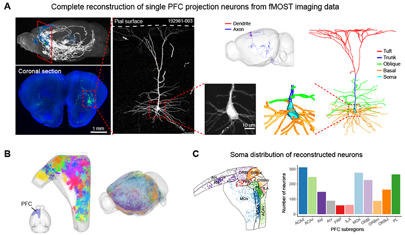
To address this question, Dr. YAN Jun's group reconstructed the dendritic morphology of 1,920 neurons (Figure 1), including 1,515 typical pyramidal neuron dendrites and 405 atypical dendrites. By quantifying the detailed dendritic morphology and subsequent clustering analysis, the researchers identified 24 types of typical pyramidal neuron dendrites as well as multiple types of atypical pyramidal neuron dendrites (Figure 2).
Next, the researchers further investigated the relationship between neuronal dendritic morphology and axonal projections and found that there was a consistent gradient of changes in dendritic morphology and axonal projections of neurons in the prefrontal cortex. For example, two types of PT neurons with different axonal projections, SCm-projecting vs. PCG-projecting PT neurons, exist in the PL-ACC region. These two types of PT neurons also exhibited different dendrite morphologies. Furthermore, by retrograde labeling and patch clamp recordings, the researchers found that these two types of neurons also differed significantly in electrophysiological properties (Figure 3).
Finally, combining the three-dimensional spatial distributions of dendrites and axons, the researchers were able to establish a network of potential connections between different neuron types in the prefrontal cortex and analyzed the nature of network connections within the same "cortical column", between ipsilateral and contralateral "cortical columns", and between different "cortical columns" on the ipsilateral hemisphere. The researchers identified a series of neuron type-specific connectivity patterns and revealed that asymmetric connections between different neuron types serve as a structural basis for information flow from superficial to deep cortical layers (Figure 4).
This work identified new dendritic types by comprehensively reconstructing and analyzing the dendritic morphology of single neurons in mouse prefrontal cortex, and systematically revealed the relationship between dendritic and axonal morphology by joint dendritic-axonal analysis. The researchers uncovered consistent gradient changes in dendritic morphology and axonal projections of PT neurons, accompanied by corresponding changes in electrophysiological properties. Finally, we used the spatial distributions of dendrites and axons to reconstruct the connectivity network between different neuronal types in the prefrontal cortex. These findings provide important clues and structural basis for further studies of the function of prefrontal cortex.
This work entitled "Single neuron analysis of dendrites and axons reveals the network organization in mouse prefrontal cortex" was published online in the journal Nature Neuroscience on May 22, 2023.
The research work was performed by Dr. GAO Le and PhD candidate LIU Sang under the supervision of Dr. YAN Jun, with important contributions from PhD candidate WANG Yanzhi , Dr. WU Qiwen , and Dr. GOU Lingfeng . The work was assisted by the research groups of Drs. XU Ninglong, SUN Yangang, YAO Haisan, XU Chun, LI Chengyu, the mouse brain mapping platform in ION, and Dr. GONG Hui's team at Huazhong University of Science and Technology. This work was funded by Ministry of Science and Technology, National Natural Science Foundation of China, Chinese Academy of Sciences, Shanghai Municipal Government, and the Lingang Laboratory.
Figure 1 Reconstruction of dendrites and axons. (A) fMOST raw image and single neuron reconstruction. (B) Dendrites and axons of reconstructed prefrontal cortex neurons (n=1920). (C) Spatial distribution of neurons in prefrontal cortex subregions.
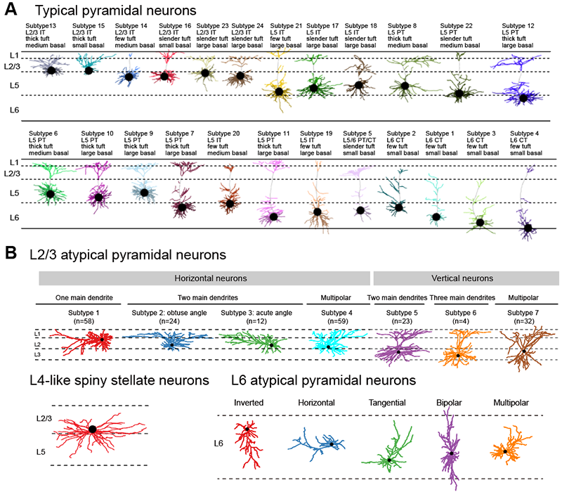
Figure 2 Dendrite types of projection neurons in the prefrontal cortex. (A) Dendrite types of 24 typical pyramidal neurons. (B) Dendrite types of L2/3 atypical pyramidal neurons, dendritic morphology of L4 spiny stellate neurons, and dendrite types of L6 atypical pyramidal neurons.
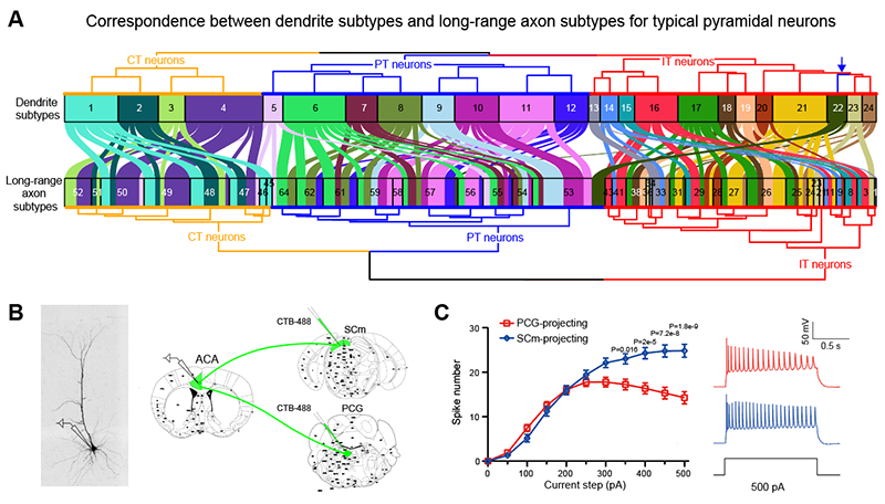
Figure 3 Correspondence between dendritic morphology, axonal projections and electrophysiological properties of typical pyramidal neurons. (A) Correspondence between dendrite subtypes and axon subtypes. (B) Schematic diagram of retrograde labeling and electrophysiological recordings of SCm- and PCG-projecting neurons. (C) Difference of spike number between SCm- and PCG- projecting under stepped injection currents.
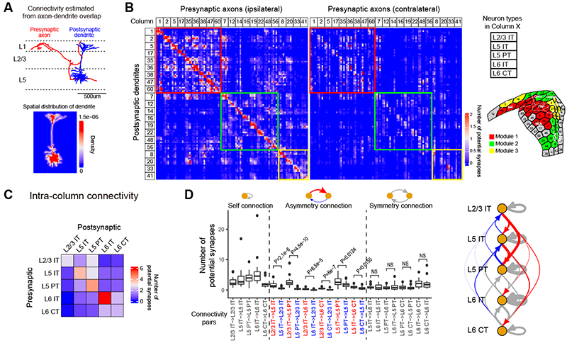
Figure 4 Construction and analysis of prefrontal cortex connectivity network based on dendritic and axonal morphology. (A) Schematic representation of the computation of the connectivity between dendrite and axon. (B) Connectivity network of prefrontal cortex neuron types. (C) Connectivity patterns of different types of neurons in the same "cortical column". (D) Analysis of the symmetry of the connections between neuron types and schematic diagram of the results.
Keywords: Relationship; Dendritic Morphology; Axonal Projections of Single Neurons
AUTHOR CONTACT:
YAN Jun
Center for Excellence in Brain Science and Intelligence Technology, Chinese Academy of Sciences, Shanghai, China.
E-mail: junyan@ion.ac.cn
 附件下载:
附件下载: 
