Time:2023-06-05
On June 1st, the journal Nature Chemistry published the Perspective article entitled: “Opportunities and challenges with hyperpolarized bioresponsive probes for functional imaging using magnetic resonance”. This is a collaborative work of Dr. Goran Angelovski from the International Center for Primate Brain Research, Center for Excellence in Brain Science and Intelligence Technology of the Chinese Academy of Sciences, Dr. Ben J. Tickner from the University of York (United Kingdom) and Dr. Gaoji Wang from Jiangsu University.
This Perspective summarizes the latest developments in the modern and exciting field of biosensor probes for hyperpolarized magnetic resonance imaging (MRI). MRI has emerged as a very important tool in biomedical research and is an essential diagnostic method in clinical radiology today. Different hyperpolarization techniques, such as dissolution dynamic nuclear polarization (d-DNP), spin exchange optical pumping (SEOP), and parahydrogen induced polarization (PHIP), can boost MRI signal intensity, and thus allow detection of wide range of biologically relevant molecules in the body that are otherwise invisible with conventional MRI methods (see Figure 1). In parallel, the chemistry of MRI contrast agents has experienced significant advances lately. This includes development of a specific class of probes known as bioresponsive MRI contrast agents which can modulate the MRI signal depending on changes in their microenvironment and thus help visualize and monitor biological processes in real time. Combined, hyperpolarized bioresponsive contrast agents can be used to make potent functional tracers and metabolites visible to MRI, and to provide valuable information about tissue metabolism and biochemistry.
To this end, the emerging field of hyperpolarized bioresponsive probes has been rapidly growing, and a wide range of hyperpolarized molecular sensors were developed very recently. In this Perspective, the authors highlight the most prominent proof-of-concept studies involving hyperpolarized biosensors produced using d-DNP, SEOP and PHIP techniques. They discussed how the amplified MRI signals of these probes can respond to biologically relevant stimuli such as target proteins, reactive oxygen species, pH or metal ions, and examined how functional MRI with these systems can allow great number of biological processes to be monitored rapidly. This work provides an exceptional outlook for further development of this methodology in advancing modern functional molecular imaging for observing physiology and pathology in real time.
The lead author of this article, Dr. Angelovski, is the head of the Laboratory of Molecular and Cellular Neuroimaging at ICPBR, CEBSIT, CAS. He has been actively involved in research focused on bioresponsive probes for functional MRI, and has published over 50 research articles, a few authoritative review papers and two book chapters in the last decade. His research group has developed a large number of MRI probes sensitive to calcium and zinc ions or amino acid neurotransmitters, including the methods for their validation by means of functional MRI in vivo.
This Perspective article published in Nature Chemistry was funded by Shanghai Municipal Government, National Natural Science Foundation of China, Jiangsu University, and Jiangsu Provincial Department of Science and Technology.
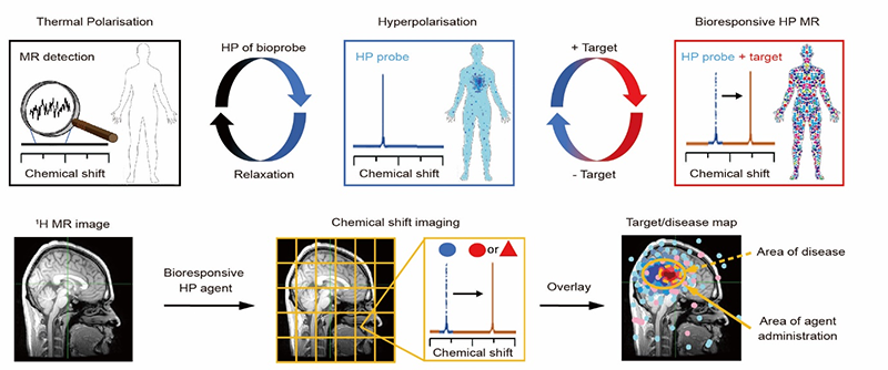
Figure 1. Perspectives for utilization of hyperpolarized bioresponsive probes for functional molecular imaging with magnetic resonance (MR). Top row: Conventional MRI suffers from low sensitivity and consequently, low concentration probes, drugs, and biomolecules are invisible to the technique (left). By employing a hyperpolarization technique, MR signals can be enhanced by a few orders of magnitude (middle). The use of probes whose MR signals can change in response to a biological target or event can reveal functional information in real time (right). Bottom row: Standard anatomical MRI images show tissue morphology (left). Functional images using a hyperpolarized bioresponsive probe can be collected using chemical shift imaging (CSI) or chemical exchange saturation transfer (CEST) approaches, which can provide spatially localized information on biological target(s) of interest (middle). Overlaying reference 1H MR images with CSI/CEST data can produce 3D maps of the distribution and concentration of biological target(s) or enzyme activity (right).
Author contact:
ANGELOVSKI Goran
International Center for Primate Brain Research, Center for Excellence in Brain Science and Intelligence Technology of the Chinese Academy of Sciences, Shanghai, China
Email: goran.angelovski@icpbr.ac.cn
 附件下载:
附件下载: 
