Time:2026-05-06
A summer in Woods Hole
It was exactly fifty years ago, in the summer of 1974, that I began my search for interesting problems in biology. I had just received my PhD from Johns Hopkins for measuring the lateral diffusion coefficient of rhodopsin in the photoreceptor membrane. Having formal training only in physics and some laboratory experience in membrane biophysics, I had no idea what I should pursue next. My thesis advisor Richard Cone suggested that I should go to the Marine Biological Laboratory (MBL) in Woods Hole, Massachusetts to find out what’s interesting in biology. Like a summer Mecca, biologists of all kinds—young and old, novice and distinguished—congregated in MBL, attending courses and conferences, giving lectures, doing experiments, or just meeting friends. From crowded lecture rooms and research laboratories to the quiet Pebble Beach and the noisy Captain Black’s café, everybody was talking about science, tossing ideas around, or gossiping about the latest news from labs. One afternoon in late August, I wandered into a new book exhibition in Lilly Hall and spotted the book On Development by J. T. Bonner. I bought the book, read it in a few days, and was totally captivated by the process of development. Bonner described in detail the potential localization mechanisms for newly synthesized materials in developing systems and extensively cited the work by Lionel Jaffe, who showed that physical forces—light, gravity, pH gradient, or electric field—could polarize rhizoid outgrowth from a fertilized fucus egg, leading to polarized root/stem development of this brown alga. Most interestingly, this polarization was accompanied by the emergence of a transcellular electric current. Considering my biophysical background, I decided to study the role of physical forces in development. Two months later, I joined Lionel Jaffe’s plant physiology lab at Purdue University.
There, I set up nerve cell cultures and attempted to measure endogenous currents around the growing neurite. The project failed completely—the current was either non-existent or too small to detect. I was soon attracted to the electric-field-induced lateral migration of membrane proteins in cultured myocytes. I began to realize that physical forces may modulate development, but chemical specificity must be the key to the intricacy of developmental processes, as exemplified by the process of synaptogenesis, where specific sets of membrane receptors and ion channels need to be localized to the synapse.
From protein mobility to synaptogenesis
When I set up my own laboratory at UC Irvine in 1976, there was a great interest in the mechanism underlying the clustering of nicotinic acetylcholine receptors (AChRs) in the developing neuromuscular junction: either they are all inserted into the postsynaptic membrane by the muscle cell, or they undergo diffusional redistribution in the plasma membrane and become trapped at the synapse. To test the latter mechanism, one needs to measure the diffusion rate of native un-liganded AChRs. I puzzled over this problem for quite some time. The solution suddenly came to me during a sleepless night—just create an asymmetry in functional AChR distribution in the myocyte membrane by local inactivation of AChRs with the irreversible blocker α-bungarotoxin and then follow the decay of the asymmetry over time to measure diffusion rate. I did the experiment the next morning, and it worked! I soon determined the diffusion coefficient of native AChRs in the myocyte membrane to be 2.6 × 10⁻⁹ cm2/s, sufficiently fast to support diffusion-mediated trapping of AChRs at postsynaptic sites.
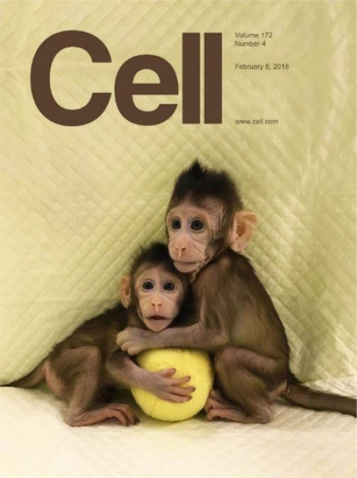
Cover of Cell issue publishing the article on the cloning of macaque monkeys by somatic cell nuclear transfer (Liu et al., 20182)
I had learned the method of culturing Xenopus spinal neurons and myocytes from Nick Spitzer at UCSD while doing my postdoctoral work at Purdue. For nearly three decades, this was the main experimental system in my laboratory for studying axon guidance and synaptogenesis to transmitter secretion and synaptic competition. Over the years, I have often been challenged on the biological relevance of our findings in cultured cells. However, I propose that any phenomenon observed in vitro is bound to be found somewhere, sometime, in vivo.
Growth cone guidance and synaptic competition
To identify important unsolved problems, the best method is to read good science books. On Development led me to my study of neuronal polarization. Dale Purves and Jeff Lichtman’s Principles of Neural Development, published in 1985, triggered many experiments in my laboratory. Many problems described in these books remain outstanding; they stand as the testimony of the progress (or the lack of progress) of the field. In the latter book, I was particularly attracted by the hypothesis of chemotactic pathfinding of nerve growth cones proposed by Ramón y Cajal and activity-dependent competition among synapses found at developing neuromuscular junctions and sympathetic ganglia.
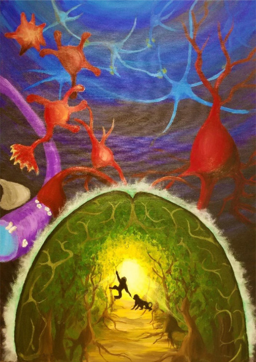
Figure viewer
Pathfinding in neurobiology from growth cone guidance to primate cognition (Oil painting by Li Ye-fei, 2018)
When nerve growth factor family of trophic factors (“neurotrophins”) and netrin and semaphorin families of guidance factors began to be identified, we developed a simple semi-quantitative in vitro method (“growth cone turning assay”) for measuring their chemotactic effects on axon growth and for studying how second messengers mediate growth cone turning behaviors. Most surprisingly, nearly all guidance factors we tested could either attract or repel the same growth cone of Xenopus spinal neurons, depending on the cyclic nucleotide (cAMP or cGMP) levels in the neuron.3 Thus, the textbook notion of chemo-attractants and chemo-repellents needs to be modified: the turning response reflects the internal state of the growth cone rather than the intrinsic property of the guidance factor. The internal state, as reflected by second messengers such as Ca2+ and cyclic nucleotides, depends on many coincident signals impinging upon the growth cone during its pathfinding in the complex extracellular matrix of developing tissues.
For activity-dependent synaptic competition, Purves and Lichtman proposed a neurotrophic hypothesis: active synapses pick up more neurotrophic factors secreted at the synapse and survive, while inactive synapses are deprived of the factors and eliminated. After moving to Columbia University, my laboratory tested this hypothesis in Xenopus nerve-muscle cultures by examining whether neurotrophic factors could modify the synaptic strength. In collaboration with Nancy Ip, we found that purified neurotrophins could rapidly potentiate developing neuromuscular synapses.4 Does electrical activity also affect developing synapses? Indeed, when a myocyte was co-innervated by two co-cultured neurons, repetitive stimulation of one neuron led to immediate suppression of synapses made by the un-stimulated neuron, whereas the stimulated synapse was unaffected. This synaptic suppression was due to a postsynaptic mechanism in the myocyte, since it could be induced simply by applying ACh pulses on the myocyte surface. Remarkably, the synapse was protected from suppression only if the presynaptic neuron was stimulated to fire concurrently (within ∼10 msec) with the applied ACh pulses.5 Now, I began to appreciate the importance of spike timing in synaptic modification.
Spike-timing-dependent plasticity
The textbook notion of activity-dependent synaptic modification was based on Hebb’s synaptic learning rule that correlated firing of pre- and postsynaptic neurons leads to synapse strengthening, while un-correlated firing weakens the synapse. However, it was unclear how closely correlated the pre- and postsynaptic spiking had to be to strengthen the synapse. After we moved to La Jolla in 1996, the neighboring laboratories of Bill Harris and Kristine Holt offered an ideal in vivo preparation to examine this problem. By precisely controlling the timing of stimulation at two different retinal ganglion cell (RGC) inputs to the same optic tectum neuron, we could determine how the strengthening and weakening of retinotectal synapses depends on the relative timing of RGC and tectum neuron spiking. The results were surprising—closely correlated pre- and postsynaptic spiking could either potentiate or depress these synapses, depending on the temporal order of spiking. By varying the timing of RGC stimulation, we charted out the time window (∼20 msec) within which pre- and postsynaptic spiking could induce either long-term potentiation (LTP, pre-before-post spiking) or depression (LTD, post-before-pre spiking) of retinotectal synapses.6 The same time window for spike-timing-dependent plasticity (STDP) was also found in cultured hippocampal neurons.7 Our results were in line with the finding of LTP/LTD induction in cortical slices reported by Henry Markram and colleagues a year earlier. The STDP with varying time windows and LTP/LTD amplitudes was later found at nearly all synapses examined.
Backpropagation of LTP and LTD
In 1986, Jeoffrey Hinton and colleagues invented the backpropagation algorithm for supervised learning in artificial neural networks (ANNs), where error signals at the output are used to modify the weight of upstream synapses reiteratively during network learning. In an article in Nature, Francis Crick expressed general excitement on the amazing power of the backpropagation algorithm but argued that this is not a plausible learning mechanism in the brain, since the information flows in neurons from input to output, not the other way around. A few years later in my UCSD laboratory, while searching for a use of our newly developed method of simultaneous whole-cell recordings from multiple interconnected neurons in culture, Crick’s comment resurfaced in my mind. We decided to test whether activity-induced synaptic changes could propagate backwards. Amazingly, we discovered that LTD induction at the output synapse of a cultured hippocampal neuron led to a substantial depression of synaptic inputs onto this neuron within a few minutes.8 In contrast, we found no forward spread of LTD to other synapses associated with the postsynaptic neuron. In my Berkeley lab a few years later, we found that activity-induced LTP and LTD at retinotectal synapses made by RGCs in the Xenopus optic tectum could backpropagate through the optic nerve to the retina, causing corresponding potentiation and depression, respectively, of bipolar cell synapses onto RGCs.9 This artificial intelligence (AI)-algorithm-inspired discovery challenges the prevailing idea that activity-dependent synaptic plasticity is localized to activated synapses, and its implication for the development and function of neural circuits in the brain remains to be explored.
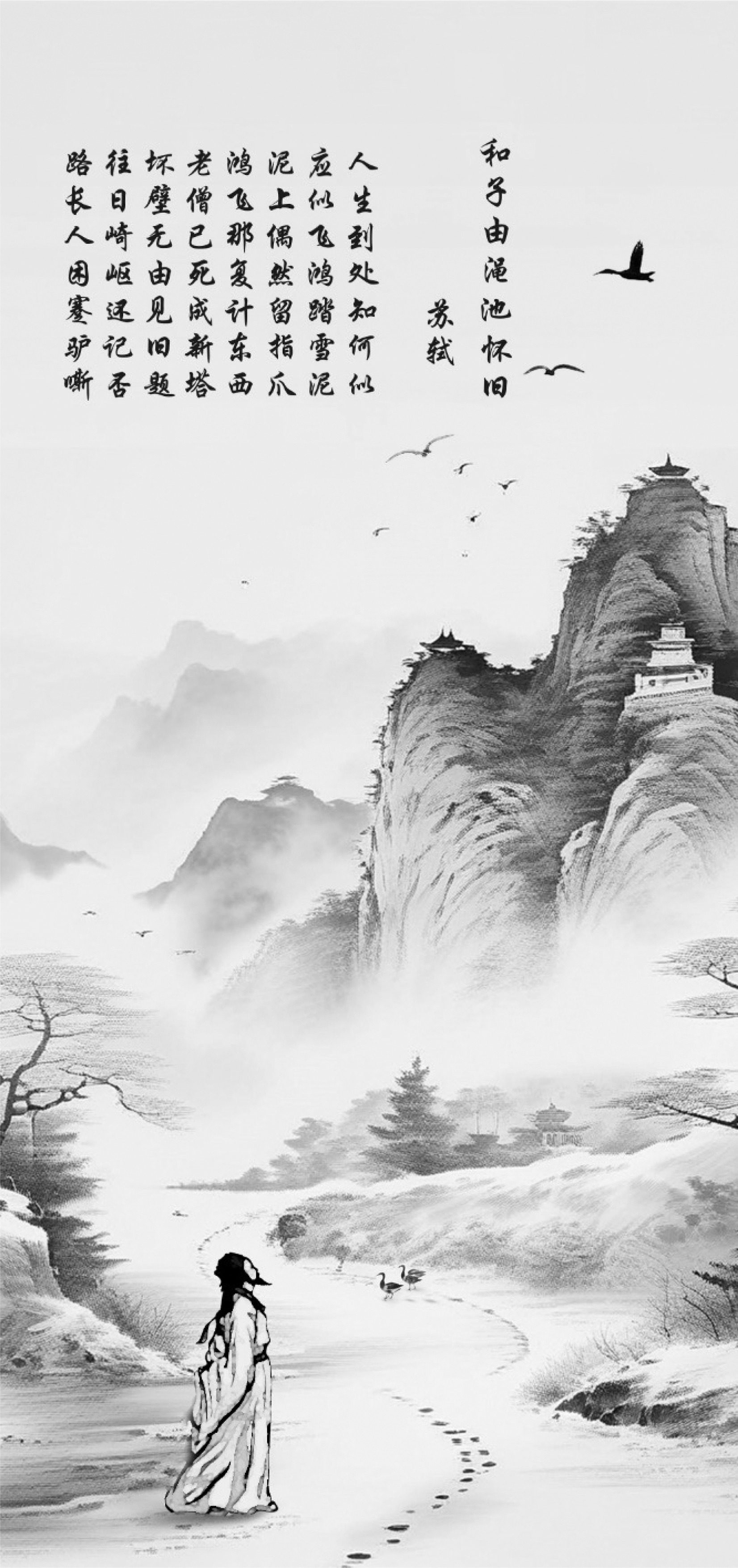
Figure viewer
Image of Song Dynasty poet Su Dung-Po lamenting the passing of his wandering life (Ink and wash painting generated by iFLY TEK’s software SparkDesk by Li Ye-fei, 2024)
In collaboration with Bo Xu and colleagues, we recently implemented this biological backpropagation in ANNs. In a three-layer spiking neural network (SNN), we induced LTP/LTD by STDP at the output synapses using expected spiking of output neurons as teaching signals and allowed potentiation/depression to backpropagate to subsets of upstream synapses in the hidden layer. This enabled the SNN to outperform other conventional backpropagation-based algorithms in several benchmark learning tasks in both accuracy and computational efficiency.10 Brain-inspired machine learning methods are likely to be increasingly relevant for developing energy-efficient sustainable AI.
Ventures in post-Cultural Revolution China
I was born in Nanjing and lived my early life in Taiwan before going to graduate school at Johns Hopkins in 1971. I made my first trip back to China in December 1981 to teach a membrane biology course at Beijing Medical College (BMC) as part of an exchange program between BMC and UC Irvine. This was followed by teaching a summer course at Tsinghua University in 1982 and a summer workshop at Nankai University in 1983, where the first patch clamp amplifier was assembled in China and used for recording single channels in cultured myocytes. The post-Cultural Revolution university campuses were full of students and faculty eager to learn but in great need of resources in teaching and research. In 1984, I became the nominal head of a newly founded department of biological sciences and biotechnology at Tsinghua University. For three years, I flew regularly to Beijing to help set up biology curricula that encouraged active student participation. My hope of contributing to research programs, however, was not realized due to the lack of funding.
In 1988, I was invited to join the Preparatory Committee for Hong Kong University of Science and Technology (HKUST) and to take charge of planning in the area of biological sciences. This led to regular trips between New York and Hong Kong for several years prior to the university opening in 1991. I recall standing on the abandoned ground originally planned for a British army base on the hilltop of Clear Water Bay, looking over the breathtaking South China Sea and imagining how a new university could rise over the hill in three years. In temporary offices in downtown Hong Kong, a large group of administrative staff was quickly assembled, and campus construction and faculty recruitment proceeded simultaneously. A group of distinguished scholars of Chinese origin were recruited from abroad as deans and department heads, who in turn planned the facility and recruited faculty in various areas. When visiting the bustling campus years later, I often regret my failure in persuading the architect to place the faculty office next door to the laboratory. I was successful in recruiting biology and biochemistry department heads and in obtaining a 16-million-dollar fund from the Hong Kong Jockey Club for developing biotechnology. Most notable was the recruitment of Nancy Ip, who had an exemplary career in HKUST. Nancy established a top-notch center for molecular neuroscience and led the development of life sciences at HKUST. Deservingly, she is now the fifth HKUST president, the first female president of a major research university in Asia.
Neuroscience in China
In 1999, I became director of a new Institute of Neuroscience (IoN) of Chinese Academy of Sciences (CAS) in Shanghai and began my 25 years of extensive involvement in Chinese neuroscience. At IoN, I introduced a new infrastructure that was more conducive to high-quality research, including appropriate research funding and salaries for investigators and staff, a simplified administrative system, a strong emphasis on quality over quantity of publications, a biennial review of research programs by an international advisory board, and a rigorous graduate student training program that meets international standards. Over one thousand PhD students have now graduated from IoN: most of them received or are receiving postdoctoral training abroad, and >100 have become independent lab heads in China and abroad. I noticed that the growth of IoN and most neuroscience institutions in China closely followed the growth of the government’s support of basic research, roughly doubling every five years for the past 25 years, and there is no sign of slowdown at this time.
As the wave of big brain projects spread across the globe, I began to participate in the discussion and planning of the China Brain Project. When the project was finally initiated in 2021, it represented a 10-year program in both basic neuroscience research on brain functions and applied neuroscience on the diagnosis and intervention of major brain diseases, as well as brain-machine intelligence technology including brain-computer interfaces, neuromodulation technologies, and brain-inspired machine learning methods and computing devices. The results of these programs are exemplified by the recent mapping of single-cell spatial transcriptomes of the entire macaque cortex11 and the generation of a live chimera monkey with high-percentage contribution of donor embryonic stem cells.12
Of mice and monkeys
The importance of using non-human primates (NHPs) as models of brain diseases has become increasingly clear in view of the striking failure in drug development using mouse disease models for preclinical efficacy tests. Although China has a rich resource of macaque monkeys, there were only a handful of laboratories doing monkey research. In 2009, I decided to build an NHP facility at IoN and recruit lab heads with NHP expertise. More than twenty IoN labs are now devoted to NHP research, ranging from reproductive biology, gene editing, and multi-omics to systems neurophysiology and cognitive neuroscience. An International Center for Primate Brain Research was inaugurated in 2019, with Nikos Logothetis and I serving as co-directors. We aim to establish NHP facilities and platforms for international collaboration in primate neurobiology. A regular summer school in primate neurobiology was also initiated in collaboration with the International Brain Research Organization.
In 2016, Zilong Qiu’s lab in IoN, working with the NHP facility director Qiang Sun, generated transgenic cynomolgus monkeys exhibiting autistic phenotypes by overexpressing the human MeCP2 gene in early embryos.13 Zhen Liu, the graduate student responsible for this work, was persuaded to stay in IoN as a postdoctoral fellow in my laboratory and work with Qiang Sun to clone macaque monkeys by somatic cell nuclear transfer (SCNT), which was then successfully applied to many mammalian species other than primates.2 Working fearlessly on a remote island in Taihu Lake, where the IoN facility was located, Liu and Sun succeeded in cloning two cynomolgus monkeys in late 2017. Cell conducted a speedy peer review and published our manuscript within one month.14 Within a year, five monkeys were cloned from skin fibroblasts of a monkey with psychotic phenotypes due to deletion of the circadian rhythm gene BMAL1 in the one-cell embryo.15 Hung-chun Chang, who initiated the BMAL1-knockout studies, is now using these BMAL1-KO monkeys to screen candidate drugs for psychosis. Zhen Liu became an IoN lab head, achieved long-term culturing of pluripotent monkey embryonic stem cells (ESCs), and produced a live chimera monkey with a high-percentage (up to 90%) contribution of donor stem cells in various tissues,13 including the brain and gonad. This paves the way for producing monkey lines with genetic modifications by editing monkey ESCs. Monkey genetics is thus moving along the path of mouse genetics, albeit at a slower pace.
Seeing thyself in the mirror
Having macaque monkeys in our IoN facility, my colleague Neng Gong and I were searching for problems that could demonstrate the usefulness of NHPs in studying higher cognitive functions. In 2014, we decided to study self-consciousness, which was then considered to be present in humans and great apes but not in monkeys. Previous studies also showed that monkeys consistently failed the standard test for self-consciousness, the mirror self-recognition (MSR) test, which requires hand-touching of the dye mark on the face in front of a mirror. Neng and Liang-tang Chang trained a group of adult rhesus macaques to understand the association between a laser spot falling on the mirror image of a monkey’s body and the somatic or proprioceptive sensation the monkey felt at the same spot on his own body. After many weeks of training, the monkeys eventually realized that the mirror images represented their own bodies, and most of them passed the MSR test.15 We now think that self-consciousness is probably innate to the animal, and our training only helped the monkey to learn that the mirror image represents its own body, an extension of its own body sense similar to the rubber hand illusion. Nevertheless, this simple experiment created a monkey model that could now be used to study the neural processes associated with self-cognition, which should be absent in the same monkey prior to the training.
 附件下载:
附件下载: 
