Time:2026-05-19
On May 15, 2026, Nature Communications published an online research article entitled “distributed neural codes of the 3D position in the marmoset frontal cortex and hippocampus”. The study identified place cells encoding the 3D position in a distributed neural network of brain areas including frontal cortex and hippocampus. This research was conducted by teams from the Center for Excellence in Brain Science and Intelligence Technology (CEBSIT), Institute of Neuroscience (ION) at Chinese Academy of Sciences with the co-corresponding authors including Dr. XU Chun Xu and Dr. LIU Cirong.
Humans and non-human primates all live and locomote in a 3D space, but the neural representation of the environment was mostly studied in a 2D space using rodents. To bridge this gap, the labs of Xu Chun, Liu Cirong and Zhao Zhengtuo at Center for Excellence in Brain Science and Intelligence Technology collaboratively developed a long-term wireless multi-channel recording system when marmosets were freely moving in a 3D space (Fig. A-D).
About 42% and 24% neurons in the frontal cortex (FC) and hippocampus (HPC) were identified as 3D position-encoding place cells, respectively (Fig. E and F). Place cells in FC and HPC exhibited similar spatial information, sparsity and stability. Compared to non-place cells, place cells exhibited stronger functional connections (Fig. G-J). Notably, their place fields were preferentially distributed in the center and border of the 3D space, and were recapitulated by an RNN-AutoEncoder model simulating the FC-HPC network. Collectively, these findings support a hypothesis that a widely distributed neural network involving frontal cortex and hippocampus constitutes a 3D positioning system in primates, in which the prefrontal cortex appears to play a more critical role. This study offers new insights into how the primate brain represent the 3D space and opens new frontiers in studying primate natural behaviors in the 3D space as well as the underlying neural mechanisms.
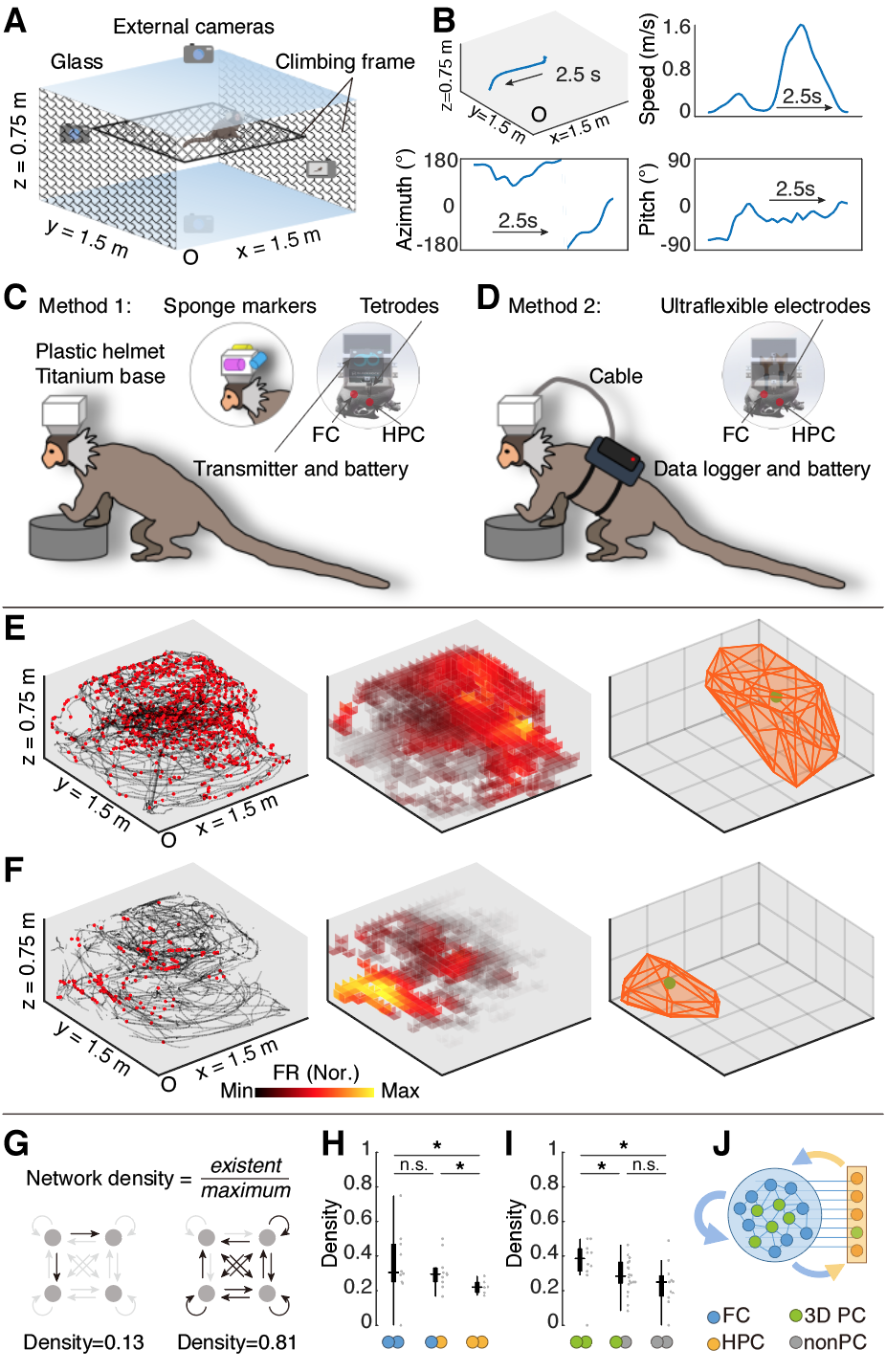
Figure legend | (A) The 3D arena and the camera-based tracking system. (B) Example passage of position, speed and head direction dynamics of marmoset locomotion. (C and D) Schematic diagram depicting wireless electrophysiological recording using 64-channle tetrodes (C) and 256-channel ultraflexible electrodes. (E and F) Examples of 3D position-encoding place cells in the frontal cortex (E) and the hippocampus (F). (G) Schematic diagram showing the quantification of functional networks. (H) Functional connections within the hippocampus are sparser than other areas. (I) Functional connections within the place cells are denser than the non-place cells. (J) Schematic diagram showing the distributed 3D position neural codes in FC-HPC networks.
Dr. Xu Chun and Dr. Liu Cirong at CEBSIT are co-corresponding authors of this work, Dr. Wei Chuanyao and Xi Jiankang at CEBSIT are co-first authors of this work. Dr. Zhao Zhengtuo, Xie Jingyi, Wang Xing, Zhu Xiaojia and Yan Haotian at CEBSIT also made contributions to this work. This study was supported by National Science and Technology Innovation 2030 Major Program, National Natural Science Foundation of China, CAS Project for Young Scientists in Basic Research, the Strategic Priority Research Program of the Chinese Academy of Sciences, Lingang Lab and the China Postdoctoral Science Foundation.
 附件下载:
附件下载: 
