Time:2026-07-06
On June 19, 2026, Nature Neuroscience published online a research article entitled “Tryptamine from wake-active monoaminergic neurons regulates sleep homeostasis.” The study was led by the research group of Dr. ZHANG Zhe at the Institute of Neuroscience, Center for Excellence in Brain Science and Intelligence Technology (CEBSIT) of the Chinese Academy of Sciences. The study reveals for the first time that cerebrospinal fluid (CSF) levels of tryptamine (TrpA) track homeostatic sleep pressure. It further demonstrates that tryptamine is produced by wake-active monoaminergic neurons and, by activating GPR139—a G-protein-coupled receptor expressed in the sleep-regulating preoptic area (POA)—raises the excitability of sleep-promoting neurons to promote sleep. The work identifies the “tryptamine–GPR139” pathway as a molecular substrate of sleep pressure and suggests it as a promising new druggable target for treating insomnia.
Wakefulness produces sleep-promoting substances, and the CSF is thought to contain signaling molecules that reflect homeostatic sleep pressure—a hypothesis proposed since the early twentieth century. Yet the identity of these molecules, the neural mechanisms that produce and sense them, and in particular whether and how the brain’s key sleep–wake regulatory neurons directly participate in this process, have long remained unresolved and await clarification.
Using targeted mass spectrometry and liquid chromatography to profile neurotransmitters and neuromodulators in the CSF, the team found that tryptamine—an intermediate metabolite of tryptophan—was markedly elevated after sleep deprivation. With a quantitative ultra-high-performance liquid chromatography (UHPLC) method they established, the researchers further confirmed that, in both nocturnal mice and diurnal miniature pigs, CSF tryptamine accumulated during wakefulness and declined after sleep, and that this change reflected the animal’s recent physical activity rather than the light-dark cycle. Intracerebroventricular infusion of tryptamine dose-dependently increased non-rapid-eye-movement (NREM) sleep and enhanced electroencephalographic slow-wave activity, displaying the characteristic phenotype of elevated sleep pressure.
To trace the source of tryptamine, the team built on a previously developed tryptophan fluorescent probe and, through site-directed mutagenesis, engineered a genetically encoded ratiometric fluorescent sensor that responds selectively to tryptamine with centisecond temporal resolution. In vivo fiber-photometry recordings with this sensor revealed that wake-active monoaminergic neurons in the locus coeruleus (LC), dorsal raphe (DR), and ventral tegmental area (VTA) release tryptamine in an activity-dependent manner during wakefulness, an effect that disappeared once synaptic release was blocked. Further experiments showed that suppressing tryptamine synthesis in the LC and DR blocked the recovery sleep rebound that follows sleep deprivation, demonstrating that tryptamine is a physiological signal linking waking activity to sleep pressure. To identify the downstream effector of tryptamine, the team performed single-cell RNA sequencing of the hypothalamic preoptic area (POA) in sleep-deprived and recovery-sleep mice and, combined with covariance analysis of immediate-early genes, pinpointed a population of inhibitory neurons that highly express the orphan receptor GPR139. Structural biology and molecular docking, together with cellular calcium-mobilization assays, confirmed that tryptamine directly binds and activates GPR139, which in turn suppresses GIRK potassium channels via the Gq/11 pathway to raise neuronal excitability. Electrophysiology and in vivo calcium recordings further showed that the excitability of POA GPR139-positive neurons rises with increasing sleep pressure.
Using Gpr139-Cre knock-in mice and CRISPR/Cas9-mediated local knockout of Gpr139 in the POA, the study demonstrated that this receptor is required for the sleep-promoting effect of tryptamine: after knockout, mice showed fragmented and lighter sleep and no longer mounted a rebound following sleep deprivation. More importantly, systemic administration of the GPR139 agonists JNJ-63533054 and TAK-041 (the latter currently in clinical trials) significantly prolonged sleep duration and deepened sleep, whereas these effects were absent in mice with Gpr139 knocked out in the POA. Human genetic analyses further found that polymorphisms in the GPR139 and AADC genes are associated with an elevated risk of sleep disorders, suggesting that targeting the “tryptamine–GPR139” pathway may represent a novel class of insomnia therapy.
In summary, the study identifies tryptamine as a previously unknown endogenous sleep-promoting molecule in the brain. As an intermediate metabolite in the synthesis of monoamine neuromodulators, it is released in an activity-dependent manner by wake-active monoaminergic neurons, accumulating during wakefulness and dissipating during sleep, and promotes sleep by activating GPR139 neurons in the preoptic area. Within the “flip-flop” framework of sleep regulation, the “tryptamine–GPR139” pathway constitutes a molecular substrate of sleep pressure that bridges waking activity and sleep, and offers a potential druggable target forsleep disorders (Fig. 1).
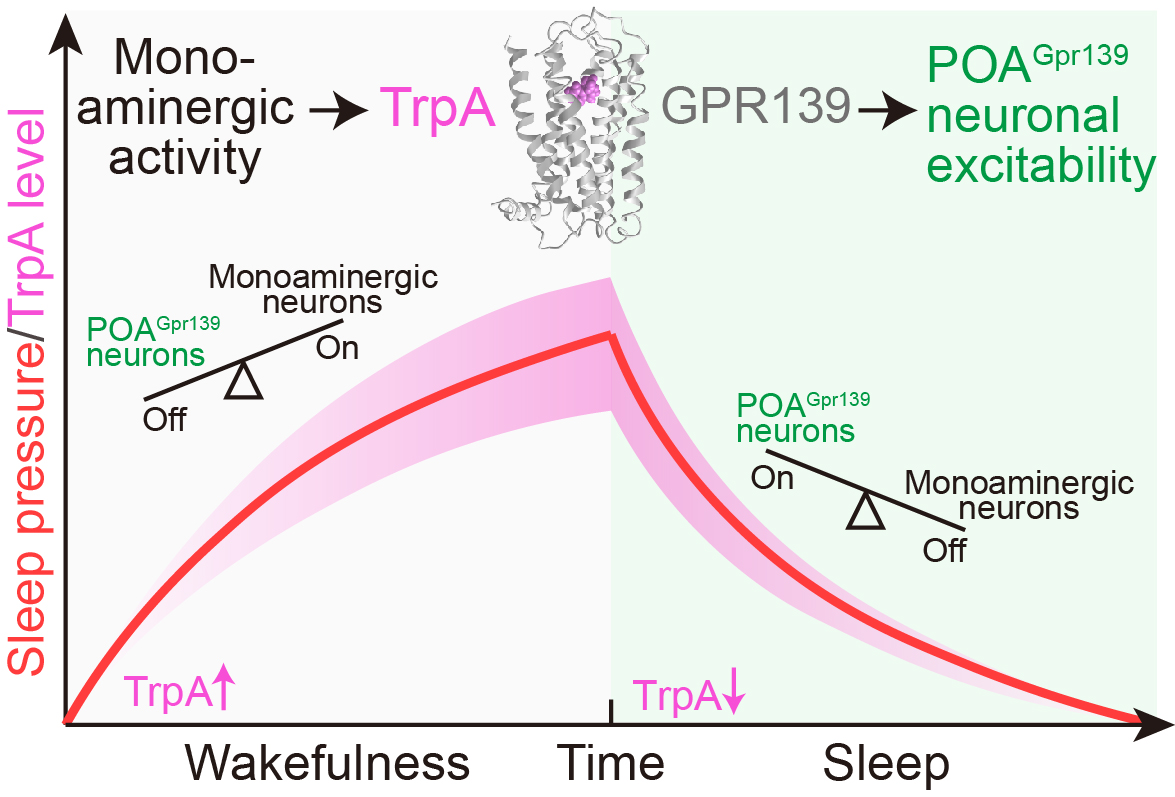
Figure 1. Schematic model of how tryptamine–GPR139 signaling encodes sleep pressure.
Dr. CAO Huateng of CEBSIT (now a young investigator at the Yiwu Research Institute of Fudan University) is the first author of the paper. Dr. ZHANG Zhe (CEBSIT), Dr. YUAN Peng (Fudan University), Prof. HU Zhi’an (Army Medical University), and Dr. MU Yu (CEBSIT) are the co-corresponding authors. Dr. WANG Kui (CEBSIT) made important contributions to the development of the sensor, Dr. ZHAO Jin provided key assistance across multiple experiments, and Dr. ZHA Zhong-Hua and Dr. LI Xiaoting (ShanghaiTech University) contributed to the structural biology experiments. The study also benefited from the valuable help of ZHANG Qian, XIU Yijin, WU Bangsheng, HUANG Shajin, CHEN Jianan, WEN Han, PAN Siwen, YANG Ke-Xin, and other collaborators. Prof. HUA Tian, Prof. LIU Zhi-Jie, and Prof. HU Ji (ShanghaiTech University), Prof. YU Jin-Tai (Fudan University), and Prof. ZHU Xiao-Na (Shanghai Jiao Tong University) provided important support. The work was supported by the technical platforms of CEBSIT and funded by the National Key Research and Development Program of China (Ministry of Science and Technology), the National Science and Technology Innovation 2030 Major Program, the National Natural Science Foundation of China, Lingang Laboratory, and the China Postdoctoral Science Foundation.
 附件下载:
附件下载: 
